Nanoscale Flow Cytometry
Extracellular Vesicle (EV) Analysis with Flow Cytometry
The Challenges of Extracellular Vesicle Analysis
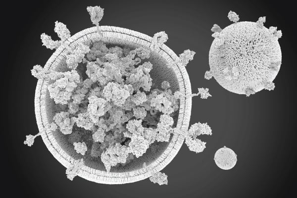
Counting, determining the size of, and characterizing extracellular vesicles (EVs) gives researchers deeper insights into cell and organ health and disease profiles. However, EVs can be challenging to analyze, owing to their small size and heterogeneity.
The need for standardization
Until now, scientists have had to use multiple techniques to accurately count, characterize and determine the size of EVs, to get the insights they need. As a result, workflows that are time-consuming and have poor repeatability are often required.
Flow Cytometry: Sensitive and Efficient Extracellular Vesicle Analysis
Flow cytometry counts, determines the size of, and characterizes EVs in one system, offering both high throughput and resolution. It can detect distinct EVs in polydisperse populations based on size scatter properties, and cargo and cellular origin based on fluorescence.
Flow Cytometry offers:
| Detailed characterization | Quality | Efficiency |
| Analyze single EVs bias-free with sensitive single particle resolution. | Reproducibly characterize heterogeneous and polydisperse samples. | Collect simultaneous multi-parametric data |
Which flow cytometry instrument is right for me?
When choosing a flow cytometry instrument, there are three key factors to consider:

| Size | Throughput | Flexibility |
| What size EV do you want to study? | How many samples do you need to run? | Do you need to have flexibility running cells and EVs? |
Our technologies make the analysis of nanoparticles by flow cytometry easy and accessible. No matter what your need is when analyzing EVs, we have it covered with the CytoFLEX Platform.
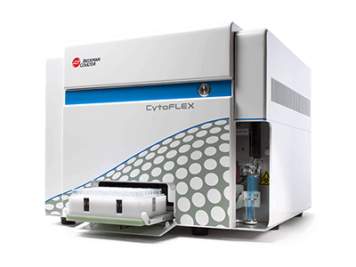
CytoFLEX Flow Cytometer
The CytoFLEX Flow Cytometer is a cell analyzer also able to detect nanoparticles down to 80 nm** thanks to its ability to measure side scatter off the violet as well as the blue laser. This allows you to characterize bigger EVs and study them within their cellular context.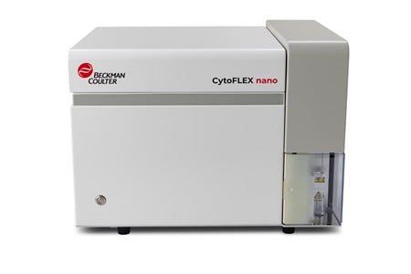
CytoFLEX nano Flow Cytometer
Built for the analysis of nanoparticles from 1 µm to at least as small as 40 nm* while simultaneously characterizing with 6 colors. The instrument has been designed to generate trustworthy results thanks to in-built automated QC processes that ensure minimal contamination and sample-to-sample carryover.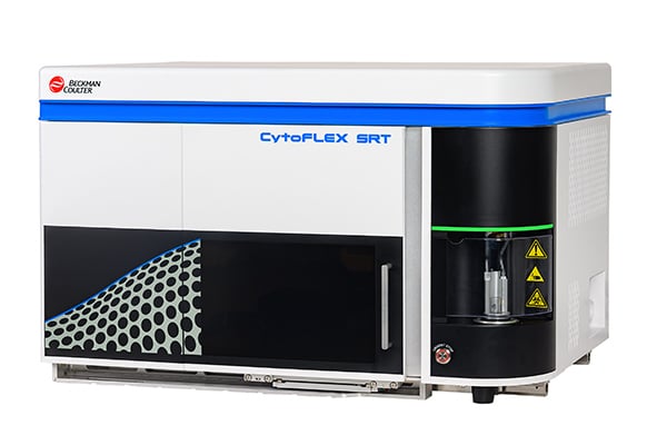
CytoFLEX SRT sorter
Drawing on the design principles of the CytoFLEX Platform of flow cytometers, this sorter efficiently handles particles smaller than 1 micron.
*Based on 40nm polystyrene beads
**Based on 80nm polystyrene beads
For Research Use Only. Not for use in diagnostic procedures.
Interested in learning how to advance your EV research?
Whether you have questions or are interested in a demo, we're here to help.


