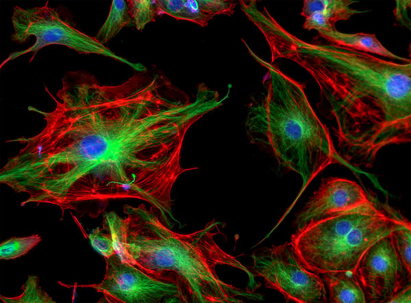
内皮细胞
内皮细胞(EC)形成内皮,一层覆盖在动脉、毛细血管和静脉等所有血管内壁上的单细胞结构。内皮细胞在血管和组织之间形成屏障,并控制物质和流体进出组织的流动。1
形成和结构
EC的形状和大小取决于其细胞特化,但它们通常薄而细长,长度约为30-50 μm。2 目前普遍认为,EC起源于胚胎和成体发育过程中的中胚层衍生的成血管细胞和血液成血管细胞。3

原始内皮细胞特化为动脉、静脉、造血和淋巴亚型的组织特异性表型,在血管发育中功能各异。研究表明,叉头盒(FOX)和ETS转录因子家族在内皮细胞的特化和建立中具有重要作用。4 此外,血管内皮生长因子(VEGF)是一种血管生成因子,可调节内皮细胞存活、增殖、分化和血管通透性,因其在许多实体肿瘤中的表达增高,目前已被证明是癌症治疗的重要候选因子。5
功能
在体内所有血管中,内皮细胞在血液和周围组织之间形成半透性屏障。作为一种重要的内分泌器官,这一单层细胞已被证明在调节血液传输、纤溶和大分子转移、免疫应答以及参与炎症应答方面发挥着重要作用。6
血管通透性
根据其性质和功能,EC可形成连续或多孔的内皮,在不同程度上支持液体和营养物质在管腔和周围组织之间运输。
血液流动
EC通过释放各种血管活性因子来启动底层平滑肌的血管扩张和血管收缩来控制血流。血管舒张因子包括一氧化氮(NO)、前列环素(PGl2)和内皮源性超极化因子(EDHF),血管收缩因子包括血栓素(TXA2)、去甲肾上腺素和内皮素-1(ET-1)。7 在炎症反应中,增加血流量是EC在增强白细胞向组织输送、增加血浆蛋白向组织渗漏和促进中性粒细胞激活方面发挥的重要作用。8
止血和血栓形成
EC对于止血和抗血栓形成方面至关重要。功能性内皮会释放抗血小板剂和抗凝剂,以防止血小板聚集和纤维蛋白形成。内皮还利用前列环素产生有效的抗血栓形成表面,通过抑制血小板聚集,保持脉管系统血浆和细胞成分流动顺畅。9当血管损伤时,EC会通过蛋白因子VIII激活血小板粘附和凝血,以及血小板聚集和纤维蛋白形成。10在伤口愈合过程中,EC释放促纤维蛋白溶解剂,通过纤维蛋白溶解降解血凝块。
在健康和疾病中的作用
血管化依赖于高度有序的发育程序,而异常生长可导致内皮功能失调。内皮细胞或血管异常通常是许多疾病的标志,包括癌症、动脉粥样硬化和其他心脏病、视网膜病变、中风、慢性肾衰竭等。内皮细胞异质性评估可以作为健康状况的有效指标;内皮功能可预测心血管疾病患者未来的心脏事件,7 研究肿瘤形成的研究人员对EC在血管生成中的作用特别感兴趣。
新兴研究
目前研究主要关注EC在许多疾病发病机制中的作用,包括病毒感染(如COVID-19)11、肝病12和肺动脉高压。13 EC细胞标志物表达的变化与内脏肥胖和胰岛素耐受性有关。14 研究还证明EC可通过调节促进生长的微环境来刺激肿瘤生长,15 这为肿瘤治疗候选靶标的选择提供了新的可能性。16
研究内皮细胞的工具
EC细胞标志物可通过流式细胞术和免疫细胞化学等免疫染色方法进行鉴定。酶联免疫吸附试验(ELISA)可量化相关的可溶性化合物,如VEGF。17 荧光色素标记溴脱氧尿苷(BRdU)掺入和CSFE染料稀释是基于流式细胞术的方法,可用于EC单细胞分析。EC培养模型可用于复制正常和病理血管的生理压力、拉伸和剪切应力。18 基因转移也正在成为一种表征EC中各个基因和蛋白质功能的有效工具。19
细胞标志物
EC表达的标志物可能取决于发育阶段和组织特异性测定。CD31,又称PECAM-1,在EC上结构性表达,而CD34在内皮祖细胞上表达,但在成熟过程中往往出现丢失。20,21 CD54 和Tek在血管EC中表达,而LYVE-1在淋巴EC表面表达。内皮细胞调控的因子也是不错的细胞标志物,包括用于血管生成的受体酪氨酸激酶VEGFR222和参与止血的糖蛋白von Willebrand 因子 (VWF)。23
特异性EC标志物概述
| 细胞标志物 | 别称 | 位置 |
|---|---|---|
| CD13 | TAPN | 细胞表面 |
| CD29 | Integrin β1 | 细胞表面 |
| CD31 | PECAM-1 | 细胞表面 |
| CD34 | 内皮祖细胞表面 | |
| CD39 | NTPDase 1, TPDase | 细胞表面 |
| CD44 | 细胞表面 | |
| CD49a | Integrin alpha-1, VLA-1, ITGA1 | 细胞表面 |
| CD54 | ICAM-1, BB2 | 细胞表面 |
| CD73 | 5'-nucleotidase, NT5E, E5NT | 细胞表面 |
| CD93 | C1qR, C1QR1, MXRA4 | 细胞表面 |
| CD105 | Endoglin, ENG, END, ORW | 细胞表面 |
| CD141 | BDCA-3, TM | 细胞表面 |
| CD143 | ACE | 细胞表面 |
| CD144 | Cadherin 5, VE-cadherin | 细胞表面 |
| CD157 | BST-1 | 细胞表面 |
| CD201 | EPCR | 细胞表面 |
| CD309 | VEGFR2, KDR, Flk-1 | 细胞表面 |
| Tek | Tie-2 | 细胞表面 |
| LYVE-1 | 细胞表面 | |
| VWF | 细胞表面 |
参考文献
1. Aman, J., Weijers, E. M., van Nieuw Amerongen, G. P., Malik, A. B., & van Hinsbergh, V. W. (2016). Using cultured endothelial cells to study endothelial barrier dysfunction: Challenges and opportunities. American journal of physiology. Lung cellular and molecular physiology, 311(2), L453–L466. https://journals.physiology.org
2. Krüger-Genge, A., Blocki, A., Franke, R. P., & Jung, F. (2019). Vascular Endothelial Cell Biology: An Update. International journal of molecular sciences, 20(18), 4411. https://www.mdpi.com
3. Tsuji-Tamura, K., Ogawa, M. Morphology regulation in vascular endothelial cells. Inflammation and Regeneration. 38, 25 (2018). https://inflammregen.biomedcentral.com
4. De Val, S., Chi, N. C., et al. (2008). Combinatorial regulation of endothelial gene expression by ets and forkhead transcription factors. Cell, 135(6), 1053–1064. https://www.cell.com
5. Duffy AM, Bouchier-Hayes DJ, Harmey JH. Vascular Endothelial Growth Factor (VEGF) and Its Role in Non-Endothelial Cells: Autocrine Signalling by VEGF. In: Madame Curie Bioscience Database [Internet]. Austin (TX): Landes Bioscience; 2000-2013. Available from: https://www.ncbi.nlm.nih.gov
6. Félétou M. (2011) The Endothelium: Part 1: Multiple Functions of the Endothelial Cells—Focus on Endothelium-Derived Vasoactive Mediators. San Rafael (CA): Morgan & Claypool Life Sciences. Available from: https://www.ncbi.nlm.nih.gov
7. Sandoo, A., van Zanten, J. J., et al. (2010). The endothelium and its role in regulating vascular tone. The open cardiovascular medicine journal, 4, 302–312. https://opencardiovascularmedicinejournal.com
8. Pober, J., Sessa, W. (2007). Evolving functions of endothelial cells in inflammation. Nat Rev Immunol 7, 803–815. https://www.nature.com
9. Rajendran, P., Rengarajan, T., et al. (2013). The vascular endothelium and human diseases. International journal of biological sciences, 9(10), 1057–1069. https://www.ijbs.com
10. Neubauer, K., Zieger, B. (2021) Endothelial cells and coagulation. Cell Tissue and Research. https://link.springer.com
11. Fosse JH, Haraldsen G, Falk K and Edelmann R (2021) Endothelial Cells in Emerging Viral Infections. Frontiers in Cardiovascular Medicine. 8:619690. https://www.frontiersin.org
12. Gracia-Sancho, J., Caparrós, E., Fernández-Iglesias, A. et al. (2021) Role of liver sinusoidal endothelial cells in liver diseases. Nature Reviews Gastroenterology Hepatology. 18, 411–431. https://www.nature.com
13. Colin E. Evans, Nicholas D. Cober, Zhiyu Dai, Duncan J. Stewart, You-Yang Zhao. (2021) European Respiratory Journal; 58(5). https://erj.ersjournals.com
14. Cifarelli, V., Appak-Baskoy, S., Peche, V.S. et al. Visceral obesity and insulin resistance associate with CD36 deletion in lymphatic endothelial cells. Nature Communications. 12, 3350 (2021). https://www.nature.com
15. Wei, C., Tang, M., Xu, Z. et al. Role of endothelial cells in the regulation of mechanical microenvironment on tumor progression. Acta Mechanica Sinica. 37, 218–228 (2021). https://link.springer.com
16. Solimando AG, Da Vià MC, Leone P, et al. (2021) Halting the vicious cycle within the multiple myeloma ecosystem: blocking JAM-A on bone marrow endothelial cells restores angiogenic homeostasis and suppresses tumor progression. Haematologica. 106(7):1943-1956. https://haematologica.org
17. Vernes JM, Meng YG. (2015). Detection and Quantification of VEGF Isoforms by ELISA. Methods Mol Biol. 1332:25-37. https://link.springer.com
18. Rosendo Estrada, Guruprasad A. Giridharan, et al. (2011). Endothelial Cell Culture Model for Replication of Physiological Profiles of Pressure, Flow, Stretch, and Shear Stress in Vitro. Analytical Chemistry. 83 (8), 3170-3177. https://pubs.acs.org
19. Zvonimir S. Katusic, Noel M. Caplice, and Karl A. Nath. (2003). Nitric Oxide Synthase Gene Transfer as a Tool to Study Biology of Endothelial Cells. Arteriosclerosis, Thrombosis, and Vascular Biology. 23:1990–1994. https://www.ahajournals.org
20. Mantovani, A., Dejana, E. (1998). Endothelium, Editor(s): Peter J. Delves, Encyclopedia of Immunology (Second Edition), Elsevier, 802-806, ISBN 9780122267659 https://www.sciencedirect.com
21. Murga M, Yao L, Tosato G. (2004). Derivation of endothelial cells from CD34- umbilical cord blood. Stem Cells. 22(3):385-95. https://academic.oup.com
22. Ferrara N, Gerber HP, LeCouter J. (2003). The biology of VEGF and its receptors. Nature Medicine. 9(6):669-76. https://www.nature.com PMID: 12778165
23. Pusztaszeri MP, Seelentag W, Bosman FT. (2006) Immunohistochemical Expression of Endothelial Markers CD31, CD34, von Willebrand Factor, and Fli-1 in Normal Human Tissues. Journal of Histochemistry & Cytochemistry. 54(4):385-395. https://journals.sagepub.com

